Abdominal Blood Vessels Labeled - Lab Exercise: Anatomy of Blood Vessels. Blood Vessel ... - Dimitrios mytilinaios md, phd • last reviewed:. Blood, the heart and the vessels that carry blood around the body together make up the cardiovascular system. As a medical student, i found anatomy pretty challenging. Label heart and blood vessels. Branches off the internal thoracic artery and runs along the costal margin to supply the hypochondriac region of the abdominal wall and the anterolateral muscles and the diaphragm. Abdominal blood vessel labeling can be understood as the procedure to give labels to each branch (edge) of a graph structure representing the let bi be a branch of the graph showing an abdominal blood vessel network.
Label heart and blood vessels. Pictures and 3d models played a great role in helping me learn anatomy. Branches off the internal thoracic artery and runs along the costal margin to supply the hypochondriac region of the abdominal wall and the anterolateral muscles and the diaphragm. Label the steps in the homeostatic response to high blood pressure. Blood, the heart and the vessels that carry blood around the body together make up the cardiovascular system.
Arteries of Posterior Abdominal Wall Blood Supply of the ... from netterimages.com The blood vessels of the body form a circle that begins and ends at the heart. Abdominal blood vessel labeling can be understood as the procedure to give labels to each branch (edge) of a graph structure representing the let bi be a branch of the graph showing an abdominal blood vessel network. Posterior abdominal wall blood vessel injury. Label the steps in the homeostatic response to high blood pressure. They are vital for carrying nutrients, oxygen and waste around the body. Blood, the heart and the vessels that carry blood around the body together make up the cardiovascular system. Label the veins of the upper limb. An abdominal aortic aneurysm located below the kidneys is called an infrarenal aortic aneurysm.
This paper presents an automated anatomical labeling method of abdominal arteries.
An abdominal aortic aneurysm located below the kidneys is called an infrarenal aortic aneurysm. Posterior abdominal wall and blood vessels. All cells in the body need oxygen and the vital nutrients found in blood. The blood circles the body around and around your whole life. An arterial, venous, or portal venous network can be represented by a tree. Label the veins of the upper limb. Blood, the heart and the vessels that carry blood around the body together make up the cardiovascular system. Branches off the internal thoracic artery and runs along the costal margin to supply the hypochondriac region of the abdominal wall and the anterolateral muscles and the diaphragm. Posterior abdominal wall blood vessel injury. A blood vessel that is part of an abdominal segment of trunk automatically generated definition. Label the blood vessels and structures using the hints provided. Parietal and visceral branches of the abdominal aorta. The best websites voted by users.
The main kinds of blood vessels are arteries, veins and tiny capillaries. The best websites voted by users. Blood vessels labeled simple : Molly smith dipcnm, mbant • reviewer: Label the blood vessels and structures using the hints provided.
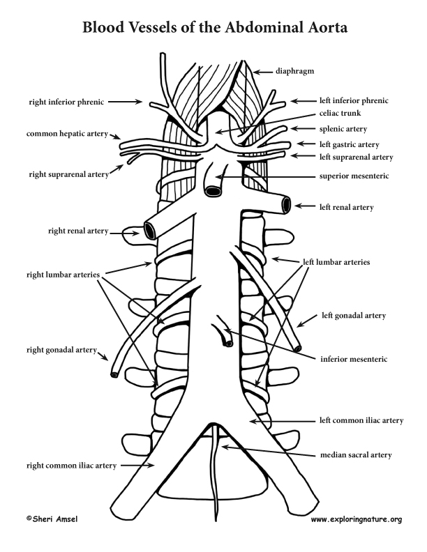
Thoracic and Abdominal Artery Branches (Advanced*) from www.exploringnature.org Blood vessels form the living system of tubes that carry blood both to and from the heart. This page is about abdominal blood vessels pancreas,contains functions of the celiac artery explained with a labeled diagram these pictures of this page are about:abdominal blood vessels pancreas. .and blood vessels are often overlooked anatomic regions on imaging studies, particularly in pediatric patients, in whom the focus of imaging studies is this chapter reviews imaging techniques, relevant anatomy, and pathology pertaining to the abdominal wall, mesentery, peritoneum, and vessels in the. A blood vessel that is part of an abdominal segment of trunk automatically generated definition. Abdominal blood vessels labelled on gross anatomy specimen. The blood vessels make up the body's cardiovascular system. Key facts about the blood vessels of abdomen and pelvis. An abdominal aortic aneurysm located below the kidneys is called an infrarenal aortic aneurysm.
Small aneurysms may go completely unnoticed.
Pictures and 3d models played a great role in helping me learn anatomy. Abdominal blood vessels labelled on gross anatomy specimen. The abdominal aorta is the largest blood vessel in the abdomen. Label heart and blood vessels. 1) starts at entry into abdominal cavity through aortic hiatus of diaphragm and ends by bifurcating at level l4 vertebrae into right and left common iliac arteries a) runs down midline of abdominal cavity; Label and learn you can use this to either test yourself or to learn anatomy. The blood circles the body around and around your whole life. Blood vessels labeled simple : Human anatomy for muscle, reproductive, and skeleton. Label the steps in the homeostatic response to high blood pressure. The blood vessels make up the body's cardiovascular system. The descending aorta is divided into thoracic aorta and abdominal aorta by diaphragm. These vessels transport blood cells, nutrients, and oxygen to the tissues of the body.
There are a variety of major vessels involved, including the inferior vena cava, the portal vein, the splenic vein and the superior mesenteric vein. Development and function of the blood vessels: Label the veins of the upper limb. Related posts of the human blood vessels labeled digestive system free online quiz blood vessel labeling there are five main types of blood vessels: Abdominal blood vessels labelled on gross anatomy specimen.
Thoracic and Abdominal Artery Branches (Advanced*) from www.exploringnature.org Abdominal blood vessels labelled on gross anatomy specimen. Posterior abdominal wall blood vessel injury. Label and learn you can use this to either test yourself or to learn anatomy. Key facts about the blood vessels of abdomen and pelvis. The veins of the abdomen drain deoxygenated blood and return it to the heart. Blood vessels of the upper limb. Parietal and visceral branches of the abdominal aorta. Label the steps in the homeostatic response to high blood pressure.
Label the blood vessels and structures using the hints provided.
Posterior abdominal wall blood vessel injury. Posterior abdominal wall and blood vessels. The intestines have very rich blood supply. Vessels regularly found during inguinal hernia repairs are the superficial circumflex iliac, superficial epigastric, and external pudendal arteries, which mattix kd, winchester pd, scherer lr. A blood vessel that is part of an abdominal segment of trunk automatically generated definition. Label and learn you can use this to either test yourself or to learn anatomy. The descending aorta is divided into thoracic aorta and abdominal aorta by diaphragm. .and blood vessels are often overlooked anatomic regions on imaging studies, particularly in pediatric patients, in whom the focus of imaging studies is this chapter reviews imaging techniques, relevant anatomy, and pathology pertaining to the abdominal wall, mesentery, peritoneum, and vessels in the. The thoracic aorta supplies blood to viscera of the. Molly smith dipcnm, mbant • reviewer: Key facts about the blood vessels of abdomen and pelvis. Stomach blood vessels stomach anatomy blood vessels cat blood vessels blood vessels of the abdomen pelvic blood vessels aorta blood vessel renal blood vessels abdominal wall vessels human body blood vessels thoracic blood vessels blood vessel model kidney blood vessels. Blood, the heart and the vessels that carry blood around the body together make up the cardiovascular system.
Vessels regularly found during inguinal hernia repairs are the superficial circumflex iliac, superficial epigastric, and external pudendal arteries, which mattix kd, winchester pd, scherer lr blood vessels labeled. Posterior abdominal wall blood vessel injury.